- Video
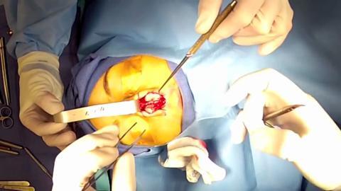
- Doctor POV: Open Rhinoplasty (Part 3)
{{ showTranscript ? 'HIDE' : 'SHOW' }} TRANSCRIPT
The nose is now been opened and what I'm grabbing is each of the lowerlateral cartilages. These white structures that you see are the left and
the right lower lateral cartilage. These essentially make up the shape of
the tip, and the shape of the base of the nose. Obviously the skin and the
nostrils are also important, but the overall shape is dictated greatly by
these cartilages.
What I'm doing now is separating the two cartilages from each other and
approaching the area called the cottel septum. That's the very front part
of the septum and it's coming into view right there. I'm spreading the
scissors on the left and the right of it and you can actually see the
structure right there.
So you can tell that they front part of the septum is closely related to
the tip cartilages and reestablishing the structural support in this area
is very important after Rhinoplasty.
What you can see is that I'm going to be measuring each of the lower
lateral cartilages. The shape, the curvature, the strength, the rigidity,
the softness, as long as the size of these cartilages dictate the shape of
the tip. What you'll also see that each cartilage is often different. It's
very rare to find perfectly symmetrical lower lateral cartilages but
classically what we like to do to narrow the tip is to measure a cephalic
trim which you'll see later on the video series; where the top part of the
cartilage is trimmed, leaving at least 6-8 millimeters of supportive
throughout.
One of the one errors of Rhinoplasty still being done today by many plastic
surgeons is removing too much of this cartilage and that's what leads to
pinched operative fake nose.
Doctor POV: Open Rhinoplasty (Part 3)
After opening the nose, Dr. Shervin Naderi separates the cartilages at the tip in order to narrow the profile and establish symmetry.